8 Immunological Diseases of the Lung

Learning objectives

  • Compare and contrast the mechanisms and manifestations of hypersensitivity pneumonitis, Goodpasture’s syndrome, systemic lupus erythematosus, rheumatoid disease, progressive systemic sclerosis, and polymyositis.


The lung is no different from any other organ in that it is susceptible to disorders of the immune system. One might argue that it is surprising that the lung does not encounter more problems, given the lung’s exposure to the environment and the myriad antigens it encounters. Before we start looking at a few specific disorders, let us quickly review the four mechanisms through which the immune system might disrupt lung tissue.


A type 1 reaction, or immediate hypersensitivity (table 8.1), is a result of overexpression of IgE (table 8.1). When an antigen binds to the overexpressed IgE on the surface of mast cells, the cell releases histamine and leukotrienes that in turn induce an inappropriate or exaggerated inflammatory response. Allergic asthma is an example of a type 1 reaction.


A type 2 reaction, or antibody-dependent cytotoxic reaction (table 8.1), is the result of a circulating antibody reacting with a component of a cell or tissue. The formation of this inappropriate immune complex results in the cell or tissue being flagged for attack by the immune system. Goodpasture’s syndrome is an example of a type 2 reaction.


A type 3 reaction, or immune complex reaction (table 8.1), is the result of an immune complex forming either locally or circulating from elsewhere and then depositing itself in tissue; the immune complex then instigates an immune system attack that involves the tissue. Pulmonary vasculitis can be caused by type 3 immune disorders.


Lastly, a type 4 reaction, or cell-mediated hypersensitivity (table 8.1), is caused by a population of hypersensitive T cells whose response to an antigen is exaggerated and leads to the proliferation of that cell population and the release of lymphokines to induce an inflammatory response. While this pattern is the same as the normal response to many infections, the magnitude of the response is inappropriate and can lead to pathological changes, such as allergic alveolitis.


Type of immune mechanism Visual Description
Type 1 - Antigen–IgE interaction
- Mast cell release of histamine, leukotrienes
- E.g., allergic asthma
Type 2 - Component of cell acts as antigen
- Antibodies bind and cell attacked
- E.g., Goodpasture's syndrome
Type 3 - A remotely or locally formed immune complex embeds into tissue
- E.g., pulmonary vasculitis
Type 4 - A sensitized T-lymphocyte responds to arrival of an antigen with proliferation and release of lymphokines
- E.g., allergic alveolitis

Table 8.1: Types of immune mechanisms involved in lung tissue injury.

With those mechanisms defined, let us look at some specific disorders.

Hypersensitivity Pneumonitis

A histological slide of lung tissue shows open air spaces but there is severe thickening of the alveolar septum.
Figure 8.1: Acute phase of hypersensitivity pneumonitis. Note presence of giant cells in the alveolar septum on the center, right-hand side of field of view.
Also called extrinsic allergic alveolitishypersensitivity pneumonitis is usually initiated by inhaled particles that are small enough to reach the alveoli and act as antigens to induce an immune response. There are many different types of particle that can be involved. The type of particle or the occupation in which the exposure occurs frequently gives its name to the condition it causes, for example farmer’s lung occurs after exposure to moldy hay and the antigen Micropolyspora foeni, bird breeder’s lung occurs with exposure to avian proteins, cheese worker’s lung occurs after inhalation and sensitization to moldy cheese. The list goes on, but the pathogenetic mechanisms are the same and the pathological/radiographic findings are indistinguishable, meaning all these diseases are considered part of the same syndrome.The time line of exposure to manifestation of symptoms, the measured serum antibody titers, and the demonstration of antibodies and inflammatory changes in the lung suggest hypersensitivity pneumonitis includes both type 3 and type 4 reactions (table 8.1).
A histological slide of lung tissue shows open airspaces in the majority of the field of view but speta appear swollen and darker lines of tissue indicate the presence of fibrotic material.
Figure 8.2: Chronic phase of hypersensitivity pneumonitis with established fibrosis.

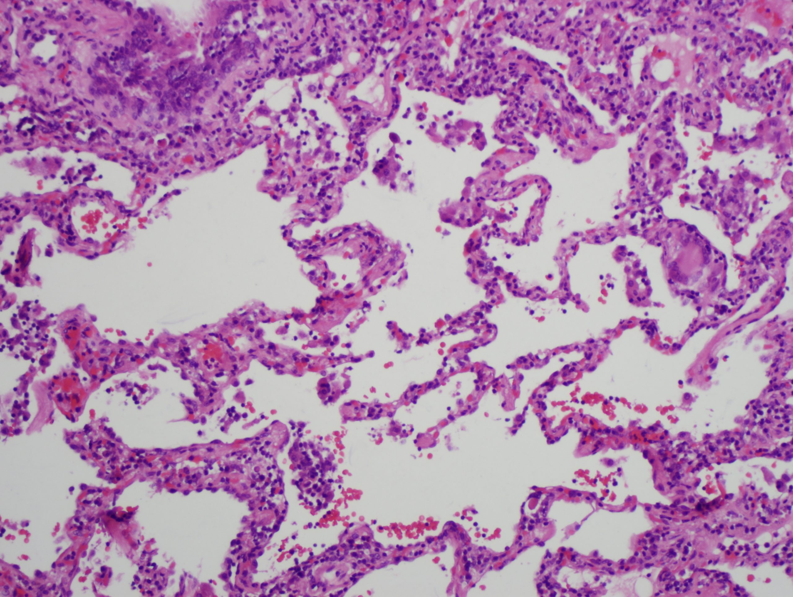
In the acute phase, lymphocytes and macrophages infiltrate the alveolar walls and loose granulomas can form. The presence of multi-nucleated giant cells (figure 8.2), are helpful in diagnosis, but more typical is a dramatic rise in lymphocyte count in BAL fluid, particularly CD8+ cells.

In the subacute phase there is evidence of interstitial thickening and the onset of fibrosis can be seen. Involvement of the bronchioles is seen with evidence of chronic bronchiolitis. The chronic form is marked by significant fibrosis (figure 8.3), to the extent it is indistinguishable from pulmonary fibrosis with distinctive fibrotic patterns and all the hallmarks of restrictive lung disease.

Clinical signs

How hypersensitivity pneumonitis presents is somewhat dependent on the form of exposure the patient had.

With brief but heavy exposure, an acute presentation of pneumonitis will present with fever, malaise, cough, and dyspnea. Physical exam confirms the fever and tachypnea and cyanosis can reflect the severity of the response. Bibasilar rales are often present, but unless a type 1 hypersensitivity arises then wheeze is usually absent (but you might note here that concurrent allergenic asthma is not beyond the realms of possibility). This acute form usually resolves within a couple of days, but can reoccur when the patient is exposed to the causal agent again.

When exposure is light but prolonged, the onset of hypersensitivity pneumonitis is more insidious and clinically challenging. The patient will describe a slowly progressive cough and developing dyspnea, also weakness and weight loss. This form of onset is common with continuous exposure to organic dust. With this longer time line the patient is not usually aware of the symptom’s relation to occupation and exposure persists until diffuse pulmonary fibrosis is established. At this point the signs and symptoms are related to respiratory insufficiency.

Goodpasture’s Syndrome

The only pulmonary disease caused by a type 2 mechanism is Goodpasture’s syndrome, which usually affects young males. It is caused by a circulating autoimmune antibody against Type IV collagen in the basement membranes of the alveoli and the renal glomeruli. Consequently the syndrome manifests as both renal and pulmonary dysfunction. The formation of an immune complex when the antibody binds to protein in the basement membrane initiates an immune response against the local tissue, and the main pulmonary manifestation is periodic hemoptysis caused by degradation of the alveolarcapillary interface.
An A-P chest x-ray shows patchy consolidation throughout all lung fields. The markings forma uniform blotchy pattern throughout the lungs.
Figure 8.3: Patchy airspace consolidation associated with Goodpasture’s syndrome.

The frequency of hemoptysis somewhat depends on the presence of other factors that affect the permeability of the lung, such as cigarette smoking or viral infection. Other manifestations include dyspnea, anemia, diffuse pulmonary infiltrate, and signs of kidney damage (hematuria as the glomerular basement membranes lose integrity).

The dyspnea no doubt occurs because of the patchy airspace consolidation that can be seen on chest x-ray as the diffuse pulmonary infiltrate (figure 8.3). In a severe episode, such as shown in this example, the patient may be hypoxic; however, distribution is often more patchy. Over the next several days the infiltrate clears but can leave a reticular pattern denoting a fibrotic reaction. Ironically, if lung transfer factor is tested the DLCO can be abnormally high because of the hemoglobin in the airspaces absorbing the carbon monoxide. The prognosis is poor with the patient usually succumbing to progressive kidney failure.

Systemic Diseases Affecting the Lung

Systemic Lupus Erythematosus (SLE)

Lupus erythematosus is a type 3 reaction. Autoantibodies are formed against cell components, particularly the nucleus and its associated proteins. The most common antibodies found are against single– or double–stranded DNA. About 70 percent of SLE patients have lung involvement that can be either acute or chronic.

Acute SLE

The acute form is referred to as lupus pneumonitis, and its signs and symptoms mimic bacterial pneumonia. It can have a rapidly progressive course with acute pleuritic chest pain and can lead to respiratory failure. The inflammation it involves can disrupt pulmonary capillaries and lead to hemorrhage. Patients with SLE are highly susceptible to infection, so diagnosis of an SLE patient should distinguish between respiratory tract infection and changes directly related to lupus. Changes in lupus come as flares followed by remission, and there are a number of triggers for flares including:

  • drugs such as tetracycline and penicillin,
  • viral infection, and
  • exhaustion or emotional stress.
An A-P chest x-ray shows diffuse reticular markings that spread from the hilar regions throughout the lungs fields to the periphery. The markings form a web-like network that have uniform density across the lungs.
Figure 8.4: Progression of diffuse fibrosis in chronic SLE.

Chronic SLE

The chronic form can progress insidiously with no symptoms or physical findings, and so it frequently it goes unrecognized until later stages of the disease. The later stages are marked with the appearance of progressive fibrosis. This is evident in the chest x-rays in figure 8.4 showing the appearance of diffuse fibrosis over a twenty-month period in a young SLE patient. The fibrosis produces reduced lung volumes and basilar atelectasis may also occur. The increased recoil of the lung can also produce an elevated and weakened diaphragm. There may also be pleural involvement with effusions arising that are normally bilateral and small.

Rheumatoid Disease

An A-P chest x-ray shows a whited-out region in the left lower quadrant of the thorax with a concave apical surface. The left lower lung is completely obscured by this feature.
Figure 8.5: Pleural effusion in a rheumatoid patient.

More famous for its effect on the joints, rheumatoid disease can also affect the lungs and pleura. Rheumatoid factors that generate rheumatoid disease continue to be investigated, but in brief, they are antibodies generated against gamma globulins. Pleural and pulmonary lesions are probably the result of local immune complex–mediated reactions associated with high levels of circulating rheumatoid factors.

Up to 50 percent of rheumatoid patients show pulmonary or pleural manifestations, with pleural involvement being more common and most frequently manifested as pleural effusion (figure 8.5). The effusate tends to have low glucose, and this finding is useful for diagnosis. Pleural and pulmonary manifestations of rheumatoid disease are more common in male patients.

A transverse CT image of the lungs shows a bright white region on the medial surface of the right lung. The region is almost circular in shape and is labelled with an arrow indicating it is a nodular lesion.
Figure 8.6: A nodular lesion (arrow) associated with rheumatoid disease.

Pulmonary involvement shows as either diffuse or nodular lesions (figure 8.6). The diffuse lesions are similar to those seen in idiopathic pulmonary fibrosis. The nodular lesions are variable in size and number and usually do not cause symptoms. They are known as necrobiotic nodules and are capable of cavitating. Bronchiolitis obliterans organizing pneumonia and bronchiectasis have also been seen in rheumatoid disease.

It should be noted here that some drugs used to treat difficult rheumatoid arthritis, such as gold preparations, methotrexate, and penicillamine, are toxic to the lung and can produce their own pulmonary lesions.

Progressive Systemic Sclerosis

Two transverse CT images of the lung are shown. The first shows diffuse, thin white markings networking throughout the lung fields shown. THe Second shows more course white markings, also throughout the lungs and these thicker markings form numerous adjacent circles that look like a honeycomb.
Figure 8.7: Fine reticular fibrosis in lower lung fields (upper CT image) progressing to honeycombing (lower CT image).

Also known as scleroderma, progressive systemic sclerosis primarily affects the blood vessels and connective tissue and likely has an autoimmune mechanism. The result is dysregulation of fibroblasts and uncontrolled collagen formation. The disease can affect many organs and tissues, and pulmonary manifestations are common.

The most common pulmonary findings are interstitial fibrosis, bronchiolar dilation, and pleural fibrosis, as well as the vascular changes that are seen in other organs. These changes produce dyspnea, cough, and basilar rales. The vascular changes can produce pulmonary hypertension that may lead to cor pulmonale.

The radiographic findings are similar to pulmonary fibrosis with the early stage of the disease showing fine reticular patterning that progresses to honeycombing in the late stage of the disease (figure 8.7). These changes are mostly found in the lower lung fields. As you might expect, the disease has restrictive characteristics along with diffusion abnormalities that produce hypoxemia during exercise.

Polymyositis

Impaired respiratory / swallowing muscle function arrow to ineffective cough arrow to poor airway clearance arrow to infection. Impaired respiratory / swallowing muscle function arrow to dysphagia arrow to aspiration arrow to infection. Drug suppressed immune response arrow to infection
Figure 8.8: The pathophysiological sequence for the most common cause of death in polymyositis.

Polymyositis is an autoimmune disease that attacks striated muscle through a cell-mediated mechanism, but can also affect other organ systems, including the lung.

There is some direct involvement with development of bronchiolitis obliterans organizing pneumonia (BOOP) and chronic interstitial pneumonitis and fibrosis. But the most frequent respiratory complications are a result of the respiratory muscles and muscles involved with swallowing becoming affected. Poor control of swallowing and inability to generate effective cough promote aspiration and retention of airway secretions. This is a recipe for bronchopneumonia—and this ends up being the most common form of death. Infection risk is often increased by the patient taking large doses of corticosteroids or immunosuppressive drugs to address the disease.

Summary

To summarize, the immunological responses to inhaled particles, localized immune responses, and systemic immunological disease can produce pulmonary manifestations that are generally related to acute and then chronic inflammatory responses. The distinguishing features and pulmonary manifestations are summarized in table 8.2.

Disorder Immunological response Pulmonary manifestations Distinguishing/associated features
Hypersensitivity pneumonitis Type 3 and 4 Fibrosis Early presence of giant cells
Goodpasture's syndrome Type 2 - Hemoptysis
- Airspace consolidation
- Associated renal disease
- Potentially high DLCO
SLE Type 3 - Acute: pneumonia-like
- Chronic: fibrosis
- Multiple organ involvement
- Butterfly rash on face
Rheumatoid disease Type 3 Nodular lesions Skeletal/joint involvement
Sclerosis Type 3 Interstitial and pleural fibrosis Associated vascular involvement
Polymyositis Type 4 Respiratory muscle weakness Inflammatory infiltration of skeletal muscle

Table 8.2: Summary of immune and systemic disorders that affect the lung.

References, Resources, and Further Reading

Text

Farzan, Sattar, with Doris L. Hunsinger and Mary L. Phillips. “Chapters 13–14.” In A Concise Handbook of Respiratory Diseases. Reston, VA: Reston Publishing Company, 1978.

Husain, Aliya N. “Chapter 15: The Lung.” In Robbins and Cotran Pathologic Basis of Disease, 9th ed., edited by Vinay Kumar, Abul K. Abbas, and John C. Aster. Philadelphia: Saunders, an imprint of Elsevier Inc., 2015.

Figures

Table 8.1: Types of immune mechanisms involved in lung tissue injury. Includes Immune mechanism – type 1 by Kindred Grey from Internet archive (CC BY 4.0), Immune mechanism – type 2 by Kindred Grey from Internet archive (CC BY 4.0), Immune mechanism – type 3 by Kindred Grey from Internet archive (CC BY 4.0), and Immune mechanism – type 4 by Kindred Grey from Internet archive (CC BY 4.0).Figure 8.1: Acute phase of hypersensitivity pneumonitis. Mutleysmith. 2012. CC BY-SA 3.0. From WikimediaCommons.

Figure 8.2: Chronic phase of hypersensitivity pneumonitis with established fibrosis. Kouranos, Vasileios, Joseph Jacob, Andrew Nicholson, and Elizabetta Renzoni. 2017. CC BY 4.0. Figure 3C from mdpi.

Figure 8.3: Patchy airspace consolidation associated with Goodpasture’s syndrome. Krzys617. 2016. CC BY-SA 3.0. From Wikidoc.

Figure 8.4: Progression of diffuse fibrosis in chronic SLE. Bickle, I., et al. 2016. CC BY-NC-SA 3.0. From https://doi.org/10.53347/rID-46209.

Figure 8.5: Pleural effusion in a 52-year old male rheumatoid patient. Pai, V., et al. 2014. CC BY-NC-SA 3.0. From https://doi.org/10.53347/rID-27112.

Figure 8.6: A nodular lesion (arrow) associated with rheumatoid disease. Chhakchhuak, Christine L., Mehdi Khosravi, and Kristine M. Lohr. 2013. CC BY 4.0. From Hindawi.

Figure 8.7: Fine reticular fibrosis in lower lung fields (upper CT image) progressing to honeycombing (lower CT image). Includes case 3 by Yang, N., Sharma, R., et al. from https://doi.org/10.53347/rID-8608 (CC BY-NC-SA 3.0) and image 1 by Gaillard, F., et al. from https://doi.org/10.53347/rID-35820 (CC BY-NC-SA 3.0).

Figure 8.8: The pathophysiological sequence for the most common cause of death in polymyositis. Grey, Kindred. 2022. CC BY 4.0. https://archive.org/details/8.9_20220203

License

Share This Book